About the project
2024
Why arthropods, why morphology, and why now?
Arthropods make up the vast majority of today’s known multicellular species. The exact number of species remains unknown, but estimates of arthropod species hover around 1,130,000, which accounts for approximately 70% of all multicellular organisms. They inhabit every part of the planet except the highest peaks and permanently frozen areas at both poles. Besides their sheer numbers, they represent a significant portion of global biomass. Renowned biologist and Pulitzer Prize winner Edward O. Wilson estimated that social insects alone, such as bees, wasps, and ants, constitute two-thirds of the biomass in the Amazon rainforest, a fact supported by more recent studies. With their vast number of species and biomass, arthropods co-create ecological processes on Earth. They are essential parts of food webs and are an untapped source of protein; as decomposers, they play a crucial role in nutrient cycling within ecosystems. Among them, we find pollinators and silk producers, as well as disease vectors and species capable of significantly reducing crop yields. One thing is certain: without arthropods, the world would not be as it is, and without them, we would not exist.
Most arthropods are overlooked in research, especially compared to our knowledge of vertebrates. Consequently, our understanding of global biodiversity and ecological processes is severely limited, and conservation efforts are less effective than they could be. Understanding the structure and function of selected arthropod species is essential for functional-ecological research, conserving functional diversity and ecosystem functions, and ultimately, for protecting ecosystem services. At a time when routine whole-genome sequencing is beginning, it’s even more crucial to understand the structure and function of organisms and their body parts, as this is the only meaningful way to understand the relationship between an organism's genotype and phenotype. Visualizing structures is the first step toward incorporating arthropods into modern functional ecology and functional genomics research.
Unfortunately, we live in an era of intense planetary change, which is associated with a massive extinction of species; many authors refer to this as the sixth mass extinction. The extinction of arthropods is no exception. In conserving the remaining life on the planet, we should consider arthropods more carefully. Moreover, numerous authors emphasize that effective nature conservation requires effectively protecting functional diversity. Visualizing the functional diversity of arthropods is our contribution to nature conservation.
Visual techniques and work plan
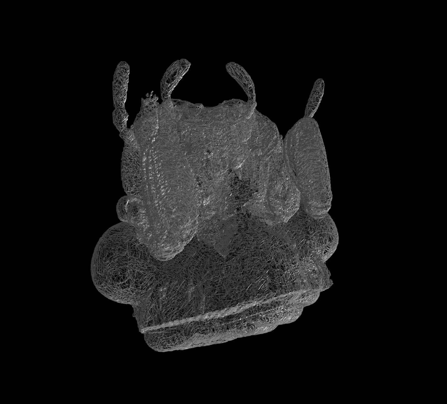
Modern visualization technologies offer an incredible insight into the structure of small organisms. The basic technique used in the 3D Atlas of Arthropods is image capture with a Neoscan N80 microtomograph. A microtomograph is a device that uses X-rays to capture a sequence of two-dimensional images of the organism from different angles, from which special computer algorithms generate detailed images of cross-sections of the entire organism in successive planes, spaced a few μm apart. From the cross-sectional images, we reconstruct a three-dimensional model of the organism, which can be observed from different angles and in various sections, providing a detailed view of the animal's structure. For the purposes of the 3D Atlas of Arthropods, the three-dimensional models are presented as 3D mesh images of external surfaces, which can be freely enlarged and rotated along all three spatial axes.
In the current phase, the project focuses on displaying the main body regions or patterns of tagmatization within all four subphyla of arthropods (chelicerates, crustaceans, myriapods, and hexapods). As new groups of organisms are added, we will gradually expand the work to include labeling and analysis of limb parts and their differentiation. The ultimate goal is a detailed anatomical analysis of arthropod external morphology, followed by the internal structure, or comparative analysis of individual organ systems, which will be integrated into a virtual arthropod collection. All results obtained within the project are freely accessible online.
Open invitation
We invite students and researchers to join the project. Students have the opportunity to develop a research or master’s thesis within the project. Through their work, they will gain experience in preparing biological samples for visualization with a microtomograph and in image reconstruction. Within the 3D Atlas of Arthropods, both students and researchers can define their own research problem in the field of functional morphology and evolution, capture images, which, with proper accreditation, will be included in the virtual collection.
Financerji
Authors
- Urban Bogataj,
- Gregor Bračko,
- Teo Delič,
- Cene Fišer,
- Žiga Fišer,
- Rok Kostanjšek,
- Rudi Verovnik,
- Miloš Vittori,
- Valerija Zakšek.
Students Vito Ham, Vesna Jurjevič, Gaj Kušar, and Adrijan Samuel Stell Pičman also participated in the project.
Authors
- Urban Bogataj,
- Gregor Bračko,
- Teo Delič,
- Cene Fišer,
- Žiga Fišer,
- Rok Kostanjšek,
- Rudi Verovnik,
- Miloš Vittori,
- Valerija Zakšek.
Students Vito Ham, Vesna Jurjevič, Gaj Kušar, and Adrijan Samuel Stell Pičman also participated in the project.